The Optical Knowledge Portal
Information – Education – Certification
Cones are one of the two types of photoreceptor cells found in the retina that respond to different wavelengths of light. The other type of photoreceptors, called rods, is responsible for dark-adapted vision. They require only a small amount of light to send useable electrical signals to the brain. By contrast, cones need sufficient illumination to work and are thus extremely sensitive to light. They are responsible for light-adapted vision and provide the basis for colour vision. There are three types of cone cells: S-cones (sensitive to blue colour), M-cones (sensitive to green colour) and L-cones (sensitive to red colour). The abbreviations stand for the different wavelength ranges to which the cone types react: short, medium and long wavelengths.

Perception of the other colours (mixed colours) is made possible by stimulating the different cone types at varying degrees. Equal stimulation of all cone types results in grey to white vision. When the field of view is exposed to extremely high light intensity, whether in part or fully, the cones become over-stimulated which results in glare. Unlike rods, cones are able to perceive much finer detail. The fovea centralis (point of sharpest vision) has a high density of cone cells and thus a high number of neuron connections to the brain. Consequently, this area within the eye is responsible for sharp central vision.
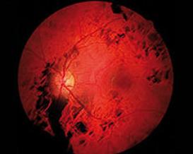
Dystrophies are genetic developmental disorders of physical structures that can lead to dysfunctions throughout life. These irreversible and degenerative changes can affect organs, tissue or even entire organisms. Cone dystrophy refers to the function loss of cones located in the area of sharpest vision. Retinal signal transmission may also be disrupted by this mutation. Since cones are responsible for colour perception, abnormal colour vision develops early on, accompanied by the gradual loss of visual acuity. Scotomas may also develop due to the fact that the visual centre contains the highest density of cones. Patients looking directly at an object may not be able to see it, because it lies within the damaged retinal area. In addition, glare sensitivity also increases as the loss of cones increases because adaption to changing lighting conditions is impaired. Other problems may also occur, such as nystagmus (‘jerking’ eyes) or strabismus (misalignment of the eyes).
As the name suggests, cone dystrophy only affects the cones. In the early stages, the cone cells in the centre of the retina die off. Vision is still possible in dim light (dusk or dawn) since rods perform this task. As the loss of photoreceptor cells progresses to the peripheral areas of the retina, rods will also be affected, which is then referred to as cone-rod dystrophy
Currently, there is no treatment available for any form of cone-rod dystrophy. Wearing sunglasses may help reduce symptoms. Blue blocking filters are ideal because they absorb UV rays and cut off a specific portion of the visible blue light subject to the selected filter. In addition, these lenses also increase contrast vision. Magnifying visual aids can also help make best use of the residual visual acuity.